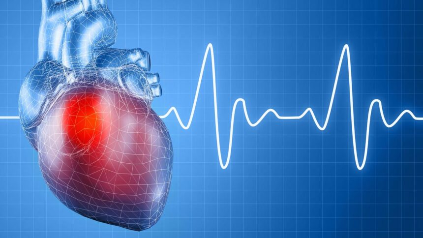
Immediately, surgeons more and more depend on 3D imaging to plan and carry out life-saving surgical procedures, particularly in conditions the place each millimeter counts, similar to aortic dissections and aneurysms. Picture used for consultant functions solely | Picture credit score: Getty Photographs/iStockphoto
Within the fashionable world of coronary heart surgical procedure, images can save lives, particularly when they’re three-dimensional. The human coronary heart and surrounding blood vessels have a posh construction, and understanding its exact form is necessary for profitable remedy. Immediately, surgeons more and more depend on 3D imaging to plan and carry out life-saving surgical procedures, particularly in conditions the place each millimeter counts, similar to aortic dissections and aneurysms.
construction
3D imaging combines knowledge from CT or MRI scans to create a lifelike digital mannequin of the center and its main blood vessels. Not like conventional 2D photos that show flat slices, these 3D fashions could be rotated, expanded, and considered from any angle. This enables docs to see what the issue seems like contained in the physique even earlier than beginning surgical procedure, permitting for cautious preparation and fewer surprises within the working room.
Some of the apparent examples of its worth is within the remedy of aortic dissection. Aortic dissection is a harmful situation wherein a tear happens in the primary artery that carries blood away from the center. Time is of the essence in these circumstances and accuracy is every little thing. Surgeons use 3D photos to see the scale, location, and extent of the laceration to assist decide the perfect surgical method. This lets you perceive how one can restore or substitute affected elements of your vessel whereas minimizing danger and plan your work nearly.
Equally, for a affected person with an aortic aneurysm (a balloon-like bulge in the identical massive artery), 3D photos can information the surgical group in deciding on the appropriately sized synthetic graft or artificial tube to strengthen the weakened vessel wall. Everybody’s physique is totally different, and so are aneurysms. 3D imaging helps customise remedy, bettering security and long-term outcomes.
Minimally invasive remedies
This similar expertise is now being utilized to minimally invasive remedies for vascular issues. When docs deal with stomach aortic aneurysms utilizing stent grafts (small wire mesh tubes positioned into the artery via a small opening within the leg), 3D imaging may also help them plan the entire thing. This enables the physician to measure the precise width, size, and curve of the artery. This ensures an ideal match of the stent-graft and reduces the prospect of leakage or repeating the process. Many hospitals create a 3D printed mannequin of a affected person’s artery earlier than surgical procedure, permitting the group to apply the process on a sensible duplicate.
However maybe probably the most thrilling growth lies in plans for transcatheter aortic valve implantation (TAVI), a brand new process to exchange broken coronary heart valves with out opening the chest. 3D imaging permits physicians to visualise a affected person’s valves, close by tissues, and blood circulation pathways with unparalleled accuracy. These detailed views will allow you to select the proper valve dimension and predict attainable issues upfront. For sufferers who’re older or too weak to endure open-heart surgical procedure, this method has made the distinction between danger and restoration.
Contained in the OT
Past planning, 3D imaging can also be beginning to play a job within the working room. Surgeons now have a display screen that shows reside reconstructed 3D photos throughout surgical procedure. This real-time steerage gives extra security and reliability, making certain gadgets are positioned in the appropriate location.
The way forward for cardiac surgical procedure is clearly shifting towards visualizing not solely the construction but additionally the dynamics of how the center and blood vessels transfer and react. The place 3D imaging analysis, planning, and execution as soon as required instinct and expertise, they’re now guided by science and readability.
For sufferers, this implies extra customized remedy, smaller incisions, sooner restoration, and a safer path to therapeutic. As expertise continues to evolve, three-dimensional imaging has turn into one of the crucial highly effective instruments obtainable to assist docs see the center because it actually is and restore it with precision.
(Dr. Shrirang Ranade is the Head and Guide of Cardiothoracic and Vascular Surgical procedure at Manipal Hospital Kalady, Pune. shrirang.ranade@manipalhospitals.com)